- Автоматизация
- Антропология
- Археология
- Архитектура
- Биология
- Ботаника
- Бухгалтерия
- Военная наука
- Генетика
- География
- Геология
- Демография
- Деревообработка
- Журналистика
- Зоология
- Изобретательство
- Информатика
- Искусство
- История
- Кинематография
- Компьютеризация
- Косметика
- Кулинария
- Культура
- Лексикология
- Лингвистика
- Литература
- Логика
- Маркетинг
- Математика
- Материаловедение
- Медицина
- Менеджмент
- Металлургия
- Метрология
- Механика
- Музыка
- Науковедение
- Образование
- Охрана Труда
- Педагогика
- Полиграфия
- Политология
- Право
- Предпринимательство
- Приборостроение
- Программирование
- Производство
- Промышленность
- Психология
- Радиосвязь
- Религия
- Риторика
- Социология
- Спорт
- Стандартизация
- Статистика
- Строительство
- Технологии
- Торговля
- Транспорт
- Фармакология
- Физика
- Физиология
- Философия
- Финансы
- Химия
- Хозяйство
- Черчение
- Экология
- Экономика
- Электроника
- Электротехника
- Энергетика
INTRAMEMBRANOUS BONE FORMATION!
INTRAMEMBRANOUS BONE FORMATION!
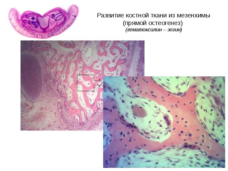
This is section of the lower jaw of the fetus!
In the uppermost small section, the germs of the teeth are visible!
On average - the bone spicules are clearly visible - they are pink! This is a small magnification!
At the lower slide you can see spicule under large magnification. The bone spicule is clearly visible. It is pink, in the center is purple osteocytes (they are already walled up in the intercellular substance). Along the edges of the spicule small violet osteoblasts are visible (they continue to secrete intercellular substance).
Large multinucleated osteoclasts are also visible (they will resorb the primary bone so that a secondary or lamellar bone appears in its place in the future)!
This is also a direct development, but schematic, taken from the atlas! You can draw the lower picture if it’s easier for you!
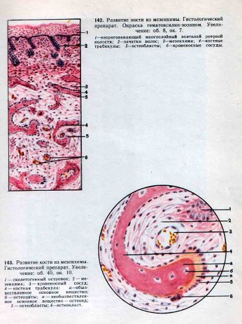
4-bone spicule; 6- osteoclast; 5- osteoblasts; b- osteosyte;
a- calcified extracellular matrix; osteoid – non-calcified ECM
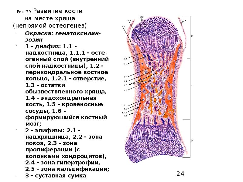
This is an endochondral bone formation! It’s immediately obvious that long tubular bones develop in this way! This is a diagram!
This is a microslide photo. endochondral bone formation!
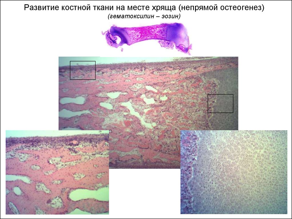
Microslide photo: long bone diaphysis(compact bone)!
|
|
|
© helpiks.su При использовании или копировании материалов прямая ссылка на сайт обязательна.
|