- Автоматизация
- Антропология
- Археология
- Архитектура
- Биология
- Ботаника
- Бухгалтерия
- Военная наука
- Генетика
- География
- Геология
- Демография
- Деревообработка
- Журналистика
- Зоология
- Изобретательство
- Информатика
- Искусство
- История
- Кинематография
- Компьютеризация
- Косметика
- Кулинария
- Культура
- Лексикология
- Лингвистика
- Литература
- Логика
- Маркетинг
- Математика
- Материаловедение
- Медицина
- Менеджмент
- Металлургия
- Метрология
- Механика
- Музыка
- Науковедение
- Образование
- Охрана Труда
- Педагогика
- Полиграфия
- Политология
- Право
- Предпринимательство
- Приборостроение
- Программирование
- Производство
- Промышленность
- Психология
- Радиосвязь
- Религия
- Риторика
- Социология
- Спорт
- Стандартизация
- Статистика
- Строительство
- Технологии
- Торговля
- Транспорт
- Фармакология
- Физика
- Физиология
- Философия
- Финансы
- Химия
- Хозяйство
- Черчение
- Экология
- Экономика
- Электроника
- Электротехника
- Энергетика
Federal State Budget Educational Establishment
Federal State Budget Educational Establishment
Of Higher education
<<Penza State University>>
Medical Institute
Course Paper
In History
<<Marcus Raichle(born 1937)>>
Student: DarjiTejaskumarDipakbhai
Group: 19ll5a
Instructor: Dr. Gavrilova Tatiana
Penza 2019-2020
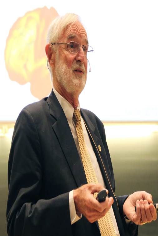
Marcus Raichle(1937)
Father of functional neuroimaging
Marcus Raichle (1937)
Marcus E. Raichle (born March 15, 1937) is an American neurologist at the Washington University School of Medicine in Saint Louis, Missouri. He is a professor in the Department of Radiology with joint appointments in Neurology, Neurobiology and Biomedical Engineering. His research over the past 40 years has focused on the nature of functional brain imaging signals arising from PET and fMRI and the application of these techniques to the study of the human brain in health and disease.[1] He received the Kavli Prize in Neuroscience “for the discovery of specialized brain networks for memory and cognition", together with Brenda Milner and John O’Keefe in 2014.
Marcus E. Raichle is a Professor of Radiology, Neurology, Anatomy and Neurobiology at the Washington University School of Medicine, St Louis. He received his bachelor’s and medical degrees from the University of Washington in Seattle. Between 1964 and 1971, he furthered his medical training at Baltimore City Hospital, Cornell Medical Centre and Johns Hopkins University, including a two-year appointment as a Major in the United States Air Force, where he worked as a Neurologist and Flight Surgeon at the USAF School of Aerospace Medicine in Texas.
He joined the faculty at Washington University as a research instructor in Neurology and Radiology in 1971, and was appointed as Professor of Neurology in 1978 and Professor of Radiology in 1979.
Marucs Raichle is known for his pioneering research in the development and use of imaging techniques, such as positron emission tomography, to identify specific areas of the brain that are involved in tasks such as seeing, hearing, reading and remembering as well as emotion. This work has allowed researchers to study the living human brain and record its function in health and disease. In addition, he and his research team have analyzed chemical receptors in the brain, investigated the physiology of major depression and anxiety, and evaluated patients at risk for stroke. He has also played a pivotal role in the development of the “default mode network” to describe resting state brain function, a concept that has become a central theme in neuroscience.
He has received many honours, including election to the Institute of Medicine in 1991 and to the National Academy of Sciences in 1996. More recently, he has received the Bristol-Myers Squibb Award for Distinguished Achievement in Neuroscience Research, the Grawemeyer Award for Psychology and the Perl-UNC Neuroscience Prize.
Noteworthy accomplishments of Marcus Raichle include the discovery of the relative independence of blood flow and oxygen consumption during changes in brain activity which provided the physiological basis of fMRI; the discovery of a default mode of brain function (i.e., organized intrinsic activity) and its signature system, the brain’s default mode network;and, the discovery that aerobic glycolysis contributes to brain function independent of oxidative phosphorylation.
His work on neuroimaging:
MRI emerged as an important imaging modality on the basis of Paul Lauterbur’s demonstration that combining classical nuclear magnetic resonance (NMR) with magnetic field gradients surrounding an object of interest it was possible to create a 3D image of the object. From this seminal observation emerged magnetic resonance imaging or MRI21. The great strength of MRI was its superb ability to image soft tissues of the body, something that CT could not do. It was an immediate success in medicine. Also it established itself as the technique for anatomical imaging of the brain, a role that it retains without rivals to this day. The idea that MRI could also image function was also present in the minds of many. The effort was to find an MRI contrast agent that was sensitive to changes in blood flow.
first attempt at using an MRI contrast agent to look at changes in brain was performed in the Martinos Laboratory of the Massachusetts General Hospital (29). They administered intravenously a paramagnetic contrast agent22 that was sensitive to changes in blood flow and were able to demonstrate that MRI could be used in this way to monitor changes in brain activity. The problem with the use of an administered contrast agent like the one used was that rapidly repeated measurements of the type being performed with PET and H2 15O were not possible.
The answer for MRI came from the Bell Laboratories and the work of Seiji Ogawa and colleagues. Seiji received his Ph.D. from Stanford in NMR working on the structure of hemoglobin. He continued to pursue research on hemoglobin at the Bell Labs. He was aware of the 1936 work of Linus Pauling on the magnetic properties of hemoglobin (30). In that work Pauling and his colleague Coryell demonstrated that deoxyhemoglobin was paramagnetic. Based on this old observation Ogawa posited that if blood flow increased more than oxygen consumption during increases in brain activity then the MRI signal would increase locally due to the reduced amount of deoxyhemoglobin present. He tested the idea by varying the concentration of oxygen in the air breathed by rodents in his MRI machine. As the concentration of oxygen was increased the dark lines representing veins in the rodent brains disappeared. From this he coined the term blood oxygen level dependent or BOLD signal which has subsequently become synonymous with fMRI (31). Because of its versatility and availability MRI has become the tool of choice for mapping the function and anatomy of the human brain as well as laboratory animals from monkeys to rodents. Four studies appearing almost simultaneous in 1991 introduced fMRI BOLD imaging in humans (32-35)
Imaging has become an increasingly important part of research in neurosciences, as well as the social sciences, and also an important face for brain research in the lay community. Using the search term “fMRI” PubMed presently finds more than 21,000 papers in the worlds literature published since the introduction of fMRI in 2001. An additional 14,000 plus papers can be attributed to PET using the search terms “PET” and “brain”. The thirst for information about brain function is universal, and imaging, for better or worse, has become a medium for the discussion.
Thank you!
|
|
|
© helpiks.su При использовании или копировании материалов прямая ссылка на сайт обязательна.
|